Benefit from individual solutions for challenging research
Your research lab or imaging facility hosts a broad range of applications. Combining imaging or optical sectioning techniques allows you to address more scientific questions with your systems. Or imaging your sample with complementary methods delivers more information about its biology. The modular concept and the broad range of imaging technologies available within ZEISS portfolio allows you to find the right combinations. Many technologies are available as field upgrades for your existing systems. You adapt your instrument to your growing needs, as new experiments become possible or new funding becomes available.
More Information
ELYRA Superresolution Combination Systems
Superresolution structured illumination (SR-SIM) and photoactivated localization microscopy (PALM) are complementary techniques with different requirements for sample preparation. SR-SIM offers full flexibility both in sample type and fluorescent label with twice the resolution of conventional light microscopy. The Nobel Prize winning, exclusively licensed PALM technology provides fluorescence microscopy with resolutions down to 20nm laterally.
Laser Scanning Confocal and ELYRA Superresolution Combined Systems
Combine LSM 880 with ELYRA for maximum flexibility in terms of resolution and three dimensional spectral imaging. You can easily switch between confocal, SR-SIM and PALM mode with the user friendly ZEN imaging software. This combined system is perfect to serve the needs of multi-user facilities with their broad range of samples, fluorophores and experimental needs.
Spinning Disk Confocal, LaserTIRF 3 and DirectFRAP Combined Systems
Whatever your live cell imaging needs, equip your microscope with a spinning disk confocal, for long term time lapse imaging. LaserTIRF 3 delivers high contrastimages of single molecules or strucutres close to membrane and cover slip. DirectFRAP allows dynamic photomanipulation studies to reveal protein dynamics. Combine all of those technologies to perform your ideal live cell experiment.
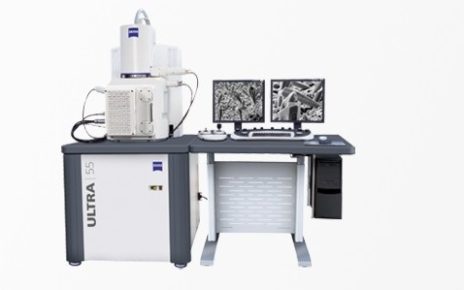
Correlate Fluorescence and Electron Microscopy Images
Image your samples with wide field, zoom – or confocal microscopy to obtain high quality, multicolor, fluorescence imaging. Then transfer the sample to your ZEISS electron microscope and image the exact same area with the Nanometer resolution. You can overlay the light and electron images for in-depth analysis of specifically tagged proteins and structures with highly detailed, nanomorphology. This gives you cutting-edge information in the context of structure and function.
Compare Microscope Techniques to Find the Right Solution
Microscopy techniques vary widely in strengths and benefits. The performance table on the next tab offers a guide to imaging techniques. Learn about the strengths and weaknesses of the different technologies for certain imaging parameters. This will allow you to identify the methods that best suit your particular types of samples or applications. If you have questions, do not hesitate to contact your application specialist from ZEISS, who has experience with multiple sample types and experimental needs and can help you to make the right decision.